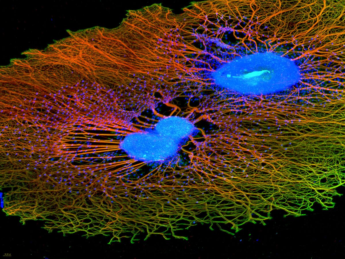
As part of our research, we develop electrochemical biosensors and matching analytical measurement modules. Find out more about our three research areas on this page.
Our three research areas
The purpose of electrochemical biosensors is to "translate" processes in biological systems into usable information. Electrodes pick up signals from a biological component and record its electrochemical activity. The biological component can consist of a biomolecule, a cell or a tissue.
The biosensor in turn consists of a system of microelectrodes and counter-electrodes to which a potential is applied. After adding a substance, the biological component changes its electrochemical properties. This change is recorded by a measuring station and then interpreted and correlated by the research team. This allows conclusions to be drawn, for example, about the effects of substances, e.g. with regard to their effectiveness or toxicity.
How a cell-based sensor is designed depends on many factors, one of which is cell and tissue type. Cardiac muscle cells behave differently than nerve cells or neuronal tissue. In addition, the material of a sensor must be biocompatible and contain various conductive, semiconductive electrode structures of different geometry and configuration.
The measuring method for which a sensor is designed also matters. If only electrochemical methods are to be used, the sensor material and the electrodes and conductor tracks do not necessarily have to be transparent. However, transparency is absolutely necessary if you also want to carry out optical measurements on high-resolution microscopes.
A new generation of sensors from our laboratory is able to combine an entire micro laboratory in a minimal area. This means that chemical syntheses, the electrophoretic purification and separation of educts and products, and the online analysis of cells and tissue can take place in the smallest of volumes on a microfluidic chip.
Drug analyzes and predictive diagnostics on human disease models in 3D format are currently already running on titer plate multi-microelectrode arrays in electronic front-end systems that we developed specifically for this purpose, with high accuracy, sensitivity and speed. Our sensors are designed, manufactured and validated in our semiconductor cleanroom, laser and electronics lab.
Many physiological processes that take place inside biological systems are subject to biophysical laws. With appropriate analysis and measurement methods, these processes can be recorded and evaluated.
The main measurement methods include cyclovoltammetry and impedance spectroscopy. Both are based on the principle of detecting electrochemical changes in a biological system and drawing conclusions from them.
In this area of our team, the source of information for our research is generated, namely cells and tissue aggregates or organotypic tissue equivalents (organoids). Using vital cell and tissue cultures, scientists are trying to record the physiology in living mode and, if necessary, to identify molecular changes that lead to a diseased condition at an early stage. These cell and tissue cultures, often based on differentiated human induced pluripotent stem cells (hiPSC), are suitable for toxicological studies and drug analysis.
In cell and tissue culture, for example, we also deal with generating pathological cell models and understanding their molecular biology and physiology. These can be tumor cells, for example, but also ischemic heart muscle cells or nerve cells that show genetically modified neuropathies. These cell models then serve as the starting point for drug research.
Three-dimensional tissue cultures represent the reality in the body much better than two-dimensional cell cultures in planar cell culture dishes. Therefore, new techniques are constantly being developed in the cell culture laboratory in order to generate corresponding 3D constructs for different cell types. This must be done individually for each cell type. Stem cells have different requirements than heart muscle cells, tumor cells need different culture conditions than retina cells.
Which culture conditions are required for which cell type is recorded in so-called Standard Operation Protocols (SOP) after a series of complex tests. Only when such an SOP could be defined is the basis for the reproducibility of an experiment given.
