Innovative technologies and complex methodological solutions form the core of all scientific cooperation at the Centre for Biotechnology and Biomedicine. These fields of expertise are clustered in technology platforms: combining infrastructures and the expertise of scientists, established method and equipment pools provide a good basis for cooperation.
Our method and equipment pools offer sophisticated technological applications with customised devices, some of them unique and specially developed in-house.
On site, the BBZ professorships that are based at BIO CITY LEIPZIG offer other research groups and biotech companies the infrastructure and expertise of nine technology lines for specific biotechnological methods and applications, such as microscopy and imaging, mass spectrometry and clean room technology.
The Following Technology Platforms Are Available at BIO CITY LEIPZIG
The Core Facility for Micro Systems Technologies of the Chair of Biochemical Cell Technology offers internal and external users access to a wide range of technologies in the field of micro systems technology.
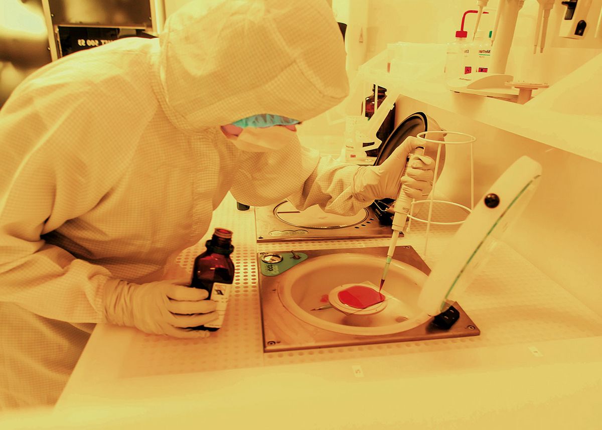
The Core Facility includes technologies for the production of microelectrode arrays, casting molds for polymer chips (PDMS-Master) and 3D printing of microfluidic platforms. Fully equipped clean rooms (class ISO5/7) with equipment for the micro systems production of electrode arrays or 3D printers and laser systems are available for this purpose.
Infrastructure and devices
The infrastructure for the production of various micro system technologies consists of three structural units:
- Cleanroom 1 for photolithographic and wet-chemical work
- Clean room 2 for additive printing techniques
- Laser system (SLE)
The cleanrooms are located on the 4th floor in the university part of the BBZ at Deutscher Platz 5 in Leipzig (rooms 1.417.1 and 1.419.1). Both cleanrooms and the equipment they contain, such as 3D printers, sandblasters, laser cutters and band saws, are assigned to the Chair of Biochemical Cell Technology. The laser system for structuring glasses is located in the Institute of Analytical Chemistry (Linnéstraße 3, Leipzig, 1st floor) and is a joint acquisition of the Professorships of Concentration Analysis (Faculty of Chemistry) and Molecular Biology and Biochemical Process Technology (BBZ).
The devices and systems integrated into the technology platform have a very wide range of specifications, so that both prototype production and the manufacture of small series are possible. Possible services and realisable quantities that can be offered are summarised in the service overview (only in german).
Comprehensive information on the infrastructure, services, utilisation models and prices of the Core Facility Micro System Technologies as well as an online form (only in german) for requesting quotations can be found on the website of the equipment centre:
Services
- Determination of the molecular weight
- Sample characterisation using LC-MS
- Confirmation of structures and structure elucidation
- Amino acid, lipid, peptide and protein analysis
- Quantification of ingredients using LC-MS/MS and LCxLC-IMS-MS/MS
- MALDI imaging analyses
This technology platform comprises five mass spectrometers with different ion sources that cover almost the entire sample spectrum of organic and biochemical analysis. With the gentle ionisation methods MALDI (matrix-assisted laser desorption/ionisation) and ESI (electrospray ionisation), the molecular weights of small organic molecules through to high-molecular biopolymers (e.g. proteins and enzymes) can be determined with high resolution (>100,000) and mass accuracy (< 1 ppm). In addition, the available MALDI-IMS-QTOF (TOF = time of flight), ESI-IMS-QTOF (Q = quadrupole), ESI-Orbitrap and ESI-Iontrap tandem mass spectrometers also offer the possibility of targeted fragmentation of molecules (collision-induced (CID) or electron transfer (ETD)). The resulting mass spectra enable the identification of "any" substances in databases and the elucidation of new or the confirmation of known structures (e.g. for amino acids, lipids, peptides and proteins). All mass spectrometers can also be coupled with analytical or nano-RP-HPLC (or -UPLC), which allows comprehensive analysis of complex samples. Tissue sections can also be analyzed using MALDI-IMS-QTOF (sample preparation on request). Highly complex samples can also be separated multi-dimensionally using orthogonal liquid chromatography (LCxLC), an ion mobility spectrometer (LCxIMS) or an electrophoretic method.
Services
- Synthesis of any peptides, consisting of the 20 proteinogenic amino acids with a chain length of 6 to 25 residues (other peptides on request; peptide quantities of 5 to 1000 mg are routinely possible)
The following modifications are possible:
- Phosphorylation on serine, threonine and tyrosine
- Acetylation or biotinylation of amino groups
- O- and N-glycosylated Ser/Thr and Asn residues
- Glycation on lysine (Amadori and Heynes peptides)
- Fluorescent dyes on amino and thiol groups
- Further modifications possible on request
In the solid phase synthesis introduced by Bruce Merrifield, peptides of any sequence and length can be synthesized. Up to a chain length of 25 residues, the synthesis is usually unproblematic, so that the peptides are generally obtained in high yield and purity. Despite the protective groups and activation methods available today, the synthesis of each peptide represents an individual challenge. About 1 - 5% of all sequences are problematic and require special synthesis strategies with optimized purification. The working group uses the Fmoc/tBu strategy, in which the base-labile fluorenylmethoxycarbonyl (Fmoc) group is used temporarily to protect the α-amino function and tert-butyl protecting groups are used to permanently protect the side chains of trifunctional amino acids. The permanent protective groups are cleaved off with trifluoroacetic acid at the end of the synthesis, whereby the peptide is simultaneously cleaved off from the solid carrier. These crude peptides can be used directly in many experiments. If higher purities are required, the peptides can be purified using reversed-phase chromatography (RP-HPLC).
Services
- Crystallization screens
- Crystallographic data acquisition
- X-ray structure analysis
- Structure interpretation
- Light scattering
- Isothermal titration calorimetry
- Microthermophoresis
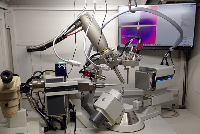
The aim of the technology platform is the crystallisation of proteins to determine the spatial structure using X-ray crystallography. Starting with a few milligrams of highly purified protein, the crystallisation conditions are first determined and optimised using a screening process with approx. 1,000 different conditions. The crystallisability of a protein sample is determined within approximately 4 to 8 weeks. Once suitable crystals have been obtained, data is collected at two measuring stations with in-house rotating anode generators or by using synchrotron radiation. In addition to determining the structure of proteins and protein-ligand complexes, molecular modelling work can be carried out for rational enzyme design or structure-based drug development. Typical questions are the optimisation of the binding affinity or the pharmacological properties of a known ligand.
In enzyme design, the planned modification of the substrate binding pocket to achieve new or broader substrate specificities is the most common goal.
Services
- Comprehensive proteome studies (2D gels and LCxLC-MS)
- 2D gel electrophoresis and 2D blots (various staining and detection techniques)
- Quantification of Western blot (various methods)
- Enzymatic cleavage in gel or in solution
- Sequence analysis of peptides with MALDI-TOF/TOF-MS or static nanoESI-QqTOF-MS (depending on the question)
- De-novo sequencing of unknown proteins and peptides
- Identification of post-translational modifications
- Saturation and minimal labelling
- Pharmacokinetic studies
The work of the technology platform comprises gel- and HPLC (UPLC)-MS-based proteome analyses, including sample preparation, processing and analysis with various software packages and databases. Gel-based proteomics generally uses 2D gel electrophoresis (IEF/SDS-PAGE; 7 cm x 10 cm up to 24 cm x 20 cm), possibly in combination with 2D blots, whereby staining methods of varying sensitivity can be used depending on the problem. Protein spots can be automatically cut out, tryptically digested in the gel (other proteases on request) and identified by mass spectrometry (MALDI-TOF/TOF or LC-ESI-MS/MS). Alternatively, an enzymatically digested sample (e.g. plasma, serum or cell digestion) can be analysed directly by mass spectrometry after one- or two-dimensional chromatography. Several thousand proteins can be identified and relatively quantified. Precise quantification can be achieved using isotope-labelled standards (synthesis in our own laboratory), for example for pharmacokinetic studies.
Services
- The techniques used in the platform include various strategies for generating overexpression vectors using genetic engineering methods, protein expression in E. coli, HEK293 and insect cells, protein analysis and finally the isolation of high-purity protein samples.
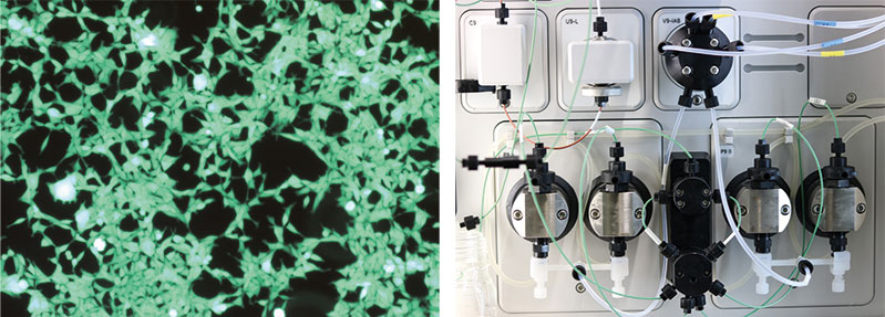
In this technology platform, proteins are made available for structural and functional analyses. While protein production was realised exclusively through bacterial expression in E. coli until a few years ago, higher expression systems are now increasingly being used for the preparation of eukaryotic proteins. The preparation of milligram quantities of eukaryotic proteins is a key technology for medical, structural biology and pharmacological projects in Leipzig. Systems for efficient overexpression in HEK293 cells have been established for this purpose. These cell lines are used in particular for the preparation of eukaryotic extracellular proteins. Expression in baculovirus insect cell systems is also utilised, particularly for cytosolic proteins and membrane proteins. For cell disruption and protein preparation, five Äkta chromatography systems are available in the laboratory for the subsequent protein purification steps.
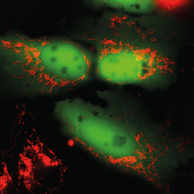
The Life Imaging technology platform provides interested parties with a Leica TCS SP5 confocal laser scanning microscope. Compared to conventional light microscopes, this microscope enables the user to capture the light emitted from a single plane of the sample. These optical sections enable sharper images and a significant improvement in 3D images of thicker samples. Another advantage is the possibility of simultaneously recording different colourants on several channels. The microscope also has a high sensitivity, which means that even very small signals can still be detected and visualised.
By using a temperature-controlled incubation chamber that can be gassed with CO2, it is also possible to observe living cells over a longer period of time. Working with the Leica TCS SP5 confocal laser microscope requires device-specific knowledge for operation. In preparation for this, the professorship offers courses by arrangement in which knowledge of the physical processes of confocal laser microscopy, sample preparation and operation of the TCS SP5 is taught.
Services
- Confocal laser scanning microscopy on living and fixed samples (life imaging is possible on living cells over longer periods of time (1-24 hours))
- Microscopy courses for confocal laser scanning microscopy
Services
- Flow cytometric analysis with up to 20 parameters simultaneously (extra- and intracellular)
- Flow cytometric sorting of up to four cell populations in parallel with up to 15 parameters simultaneously
- Performance of flow cytometry as a multi-user unit: measurements are performed by the user themselves after thorough familiarisation by core unit personnel (all measures for device maintenance, preparation of the devices, especially the cell sorter, and measurement are performed by the responsible personnel)
The Core Unit Flow Cytometry (CUDZ) provides two high-performance flow cytometers for analysing and sorting biological substances (cells, bacteria, unicellular parasites, etc.). With the help of the LSRFortessa analyser, it is possible to carry out cell characterisation at a very high level. Due to the large number of parameters that can be analysed simultaneously, it is possible, for example, to investigate cell activation states and measure cytokine profiles at the same time. Intracellular staining is ideal for this purpose. Equipped with a UV laser, analysis with DNA dyes for the characterisation of stem cells (side population) and the measurement of calcium currents is also possible. By using special staining techniques, such as phospho-flow, intracellular signalling pathways can be investigated. Far fewer cells are required for this than for conventional analyses using Western blots.
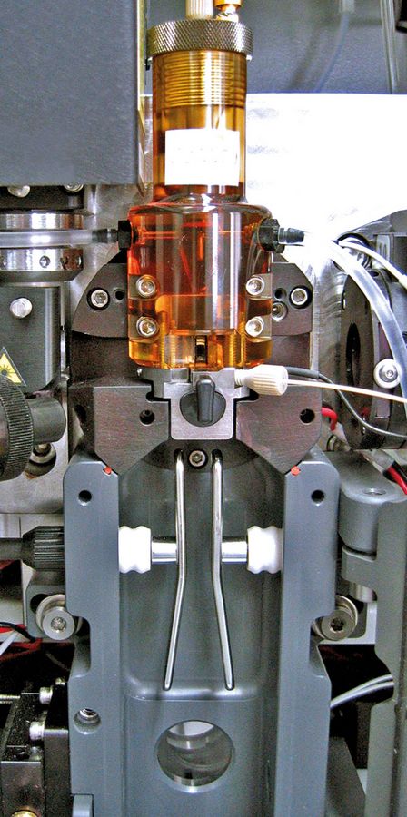
The FACSAria III high-speed cell sorter is used for fast and gentle sorting of biological substances (cells, bacteria, unicellular parasites, etc.). The samples can be sorted sterile so that the sorted cells can then be used for further cultivation experiments. Single cell sorting is also possible, e.g. for RNASeq analyses. A maximum of four different fractions can be sorted in one run. The devices are located in an S2 area (in accordance with the Infection Protection and Genetic Engineering Act). It is therefore also possible to analyse/sort infectious samples and to examine genetically modified organisms.
Further Technological Support From the BBZ Members
In addition to the method and equipment pools at BIO CITY LEIPZIG, our members have also developed a number of technological services so that other research institutions and companies from the life science sector can benefit from their expertise in the technologies developed in the Centre’s research network. These offers are supervised by the Department of Research Services.
